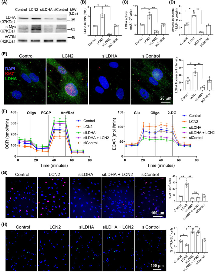
FIGURE 4.

LCN2 induced LDHA expression promotes proliferation and decreases apoptosis in PASMCs. (A–D), In vitro cultured human PASMCs were treated separately with LCN2 (2 nM), LDHA siRNA (siLDHA, 0.1 μM), control siRNA (siControl, 0.1 μM) or saline for 24 h. The protein levels of LDHA and c‐Myc, LDHA mRNA levels, LDHA activity and intracellular lactate levels were measured. (E), Representative images of immunofluorescent staining for LDHA (green), Ki67 (red) and DAPI (blue) to show intracellular LDHA expression. (F), Extracellular acidification rate (ECAR) and oxygen consumption rate (OCR) were measured to evaluate the glycolysis and mitochondrial respiration. (G and H), Representative images and quantification analyses of cell proliferation using Ki67 staining and cell apoptosis using TUNEL assay were shown. Results are expressed as mean ± SE; n = 4, independent experiments. Statistical significance was determined by Kruskal–Wallis test. * p < 0.05 and ** p < 0.01; ns p > 0.05.
