Figure 1.
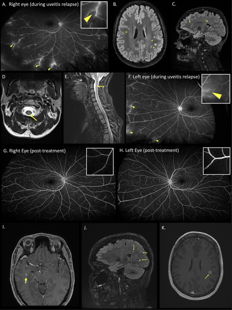
Case 1 (A) fluorescein angiography of the right eye during a relapse of uveitis. Arteriovenous phase showing leakage of fluorescein dye in peripheral retinal veins (arrowheads and inset) consistent with retinal periphlebitis. (B-C) Axial and Sagittal T2 FLAIR MRI sequences of the brain showing multifocal white matter hyperintense lesions in a distribution typical of multiple sclerosis (arrowheads). (D-E) Axial and sagittal T2 MRI sequences of the spine showing an ill-defined left posterior hyperintense lesion at the level of C2 (arrows). (F) Fluorescein angiography of the left eye upon relapse after treatment interruption of 5 months, showing retinal periphlebitis (arrowheads and inset). (G-H). Progress wide-field fluorescein angiography (early arteriovenous phase) performed 7 months after restarting treatment, showing resolution of retinal periphlebitis (inset). Case 2 (I) Axial T1 post-contrast MRI brain showing a gadolinium-enhancing lesion (arrow) in the right anterior temporal horn white matter. (J) Sagittal T2 FLAIR MRI brain showing non-enhancing juxtacortical white matter lesions (arrows). (K) 4 months later, axial T2 post-contrast MRI brain showed a new enhancing lesion in the left inferior frontoparietal lobe (arrow).

