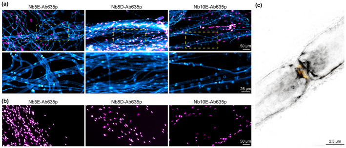
FIGURE 6.

Specific staining of mCNPase using fluorescently labelled nanobodies. (a) Epifluorescence images of teased sciatic nerves from wild‐type mice stained with Nb5E, Nb8D, and Nb10E displayed in cyan, nuclei stained with DAPI displayed in magenta. (b) Teased sciatic nerves from mice lacking CNPase display no staining (cyan) and nuclei in magenta. Images were acquired with the same acquisition settings and are equally scaled for display and direct comparison. (c) 2‐Colour 2D STED image using Nb8D‐Ab635p (grey) and voltage‐gated sodium channels (Nav1.6; yellow).
