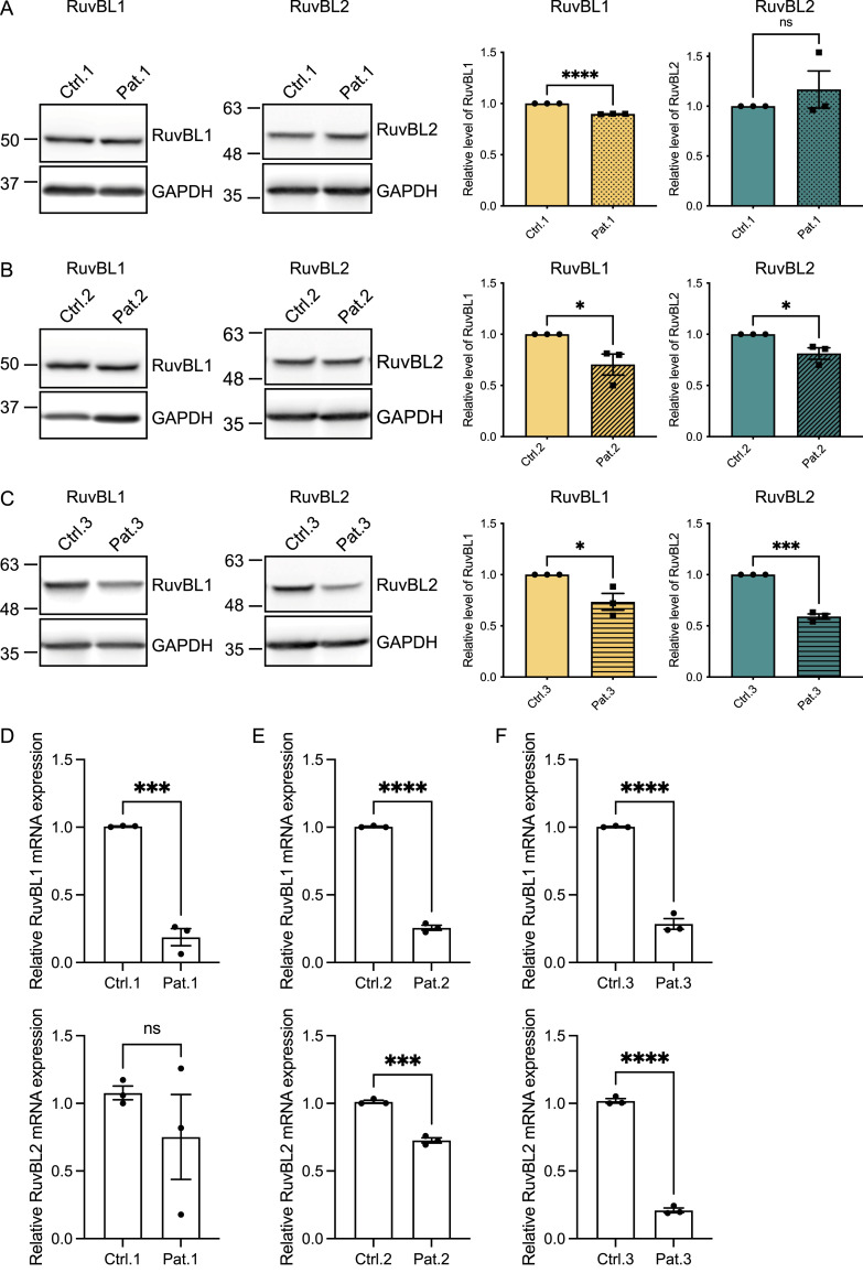
Figure 2. C9ALS/FTD patient cells have reduced levels of RuvBL proteins.
(A, B, C) RuvBL1 (left immunoblot) and RuvBL2 (right immunoblot) protein levels from 3 C9orf72-ALS/FTD patient iNPC lines and their age and sex-matched controls were determined by immunoblot. Levels of RuvBL1 (left graph) and RuvBL2 (right graph) were normalised to GAPDH and are shown relative to the age and sex-matched control (mean ± SEM; unpaired t test: *P ≤ 0.05, ***P ≤ 0.001, ****P ≤ 0.0001, ns, non-significant; N = 3 independent experiments). (D, E, F) Expression of RuvBL1 (upper graphs) and RuvBL2 (lower graphs) transcripts were quantified by RT-qPCR using 18S as a housekeeping gene (mean ± SEM, N = 3 independent experiments; unpaired t test: **P ≤ 0.005, ***P < 0.001, ****P < 0.0001).

