Figure 2.
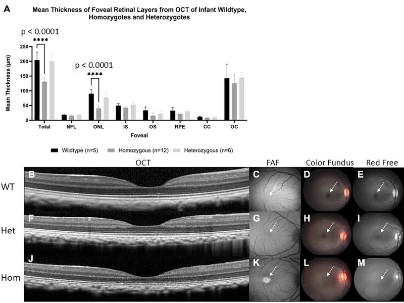
Homozygous infants were shown to have reduced foveal ONL thickness when compared to wildtype and heterozygous infant macaques. (A) Mean ± SEM thickness of retinal layers from spectral domain optical coherence tomography (SD-OCT) of infant wildtype (n = 5), homozygotes (n = 12), and heterozygotes (n = 6) macaques. Foveal ONL thinning is observed in homozygous infant rhesus macaques. Two-way ANOVA revealed a significant interaction between genotype and retinal layer thickness at total foveal thickness and at foveal ONL thickness, F (14, 160) = 7.4, P < 0.0001. Further post hoc analysis revealed a significant decrease (P < 0.0001) in homozygote infants (130.9 ± 3.6 and 40.5 ± 2.3) when compared to infant wildtype (204.3 ± 12.0 and 89.9 ± 6.5) at total foveal thickness and at foveal ONL thickness. No statistical significance was found between infant wildtype and heterozygotes. Images show a comparison of macular regions in infant wildtype, heterozygous, and homozygous primates aged 3 to 7 months. SD-OCT scans depict normal foveal appearance in wildtype (B) and heterozygous (F) primates but foveal thinning in homozygous infant (J). The macular area is marked with white arrows directing attention to macular differences among wildtype, heterozygous, and homozygous primates. Fundus autofluorescence depicting normal macular appearance in wildtype (C) and heterozygous (G) infants, but prominent foveal hyperautofluorescence is seen in homozygous infants (K). Color fundus shows normal macular appearance in wildtype (D) and heterozygous (H) but absent foveal light reflex in homozygous infants (L). Red free fundus photography showing normal foveal light reflection in wildtype (E) and heterozygous (I) but absent foveal light reflex in homozygous macaques (M).

