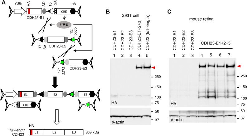
Figure 8.
CRE-lox-mediated reconstitution of CDH23 delivered by tripartite AAV vectors. (A) Schematic of the CDH23 reconstitution using tripartite AAV-CDH23 vectors. The CDH23 CDS (10 065 bp) was split into three pieces (E1: 2176 bp, E2: 4077 bp, and E3: 3812 bp), and the CRE gene was included in the 5′ (E1) vector for self-inactivation after recombination. A T2A “self-cleaving” peptide was used for CRE expression. An HA tag (red) was added to the N-terminus of CDH23 for detection (after the signal peptide). (B) Production of full-length CDH23 proteins (red arrowhead) by CRE-lox-mediated recombination in 293T cells. HEK293T cells were transduced with tripartite AAV2/2-CDH23 vectors (MOI: 3 × 10⁴ GC/cell of each vector), and cell lysates were subjected to SDS-PAGE followed by immunoblotting with HA antibodies. Lane 1: AAV-CDH23-E1 only, lane 2: AAV-CDH23-E2 only, lane 3: AAV-CDH23-E3 only, lane 4: AAV-CDH23-E1, E2, and E3 co-transduced, and lane 5: pSS-HA-CDH23-SF plasmid transfected (full-length; positive control). β-actin was used as a loading control. (C) Expression of CDH23 from the tripartite AAV-CDH23 vectors in mouse retinas. Tripartite AAV-CDH23 vectors were subretinally administered to wild-type mice as indicated (lane 1: E1 vector alone, lane 2: E2 vector alone, lane 3: E3 vector alone, lanes 4–7: E1, E2, and E3 co-injected) at the dose of 3 × 10⁹ GC per vector (n = 3–4 per vector or vector set). Treated eyes were collected 3 weeks post-injection and retinal protein extracts were subjected to SDS-PAGE and immunoblotting. Each lane represents an individual eye.

