Figure 5.
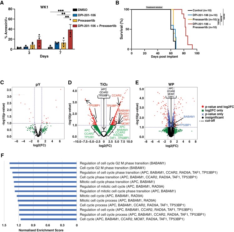
DPI-201-106 validation in a WK1 glioblastoma model. (A) AnnexinV apoptosis analysis was examined in WK1 cells treated with 10 µM DPI-201-106 after 3 and 7 days. Data shown are mean ± SD, and statistical significance was determined with a 2-ANOVA and Tukey’s multiple comparison test. (B) Kaplan-Meier survival curve of WK1-bearing mice treated with vehicle, DPI-201-106, prexasertib, or a combination of DPI-201-106 and prexasertib. Data are presented as the percentage survival of mice in each group where statistical significance was determined between the combination treatment and the other treatment groups with a log-rank (Mantel-Cox) test. Mass spectrometry analysis was performed on WK1 cells treated with DPI-201-106 for 24 h and volcano plots for (C) tyrosine phosphorylated peptides, (D) serine/threonine phosphorylated peptides, and (E) whole protein peptides were generated using Python. (F) Normalized enrichment scores of significant gene sets identified by GSEA of serine/threonine phosphorylated peptides in DPI-201-106-treated WK1 cells. **p < .01, ***P < .001, ****P < .0001. pY, tyrosine phosphorylated; TiO2, serine/threonine phosphorylated; WP, whole protein.

