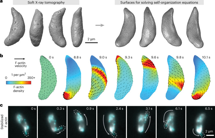
Fig. 3. Stable actin filaments circle the Toxoplasma cell.
a, Soft X-ray tomograms of cryo-fixed extracellular Toxoplasma gondii tachyzoites were used to generate triangle-meshed surfaces on which to solve our actin self-organization theoretical model. b, For stable filaments, solving the model constrained to Toxoplasma’s surface geometry predicts recirculating actin patches. c, In experiments, actin filaments briefly stabilized with jasplakinolide can circle around the cell. Cyan and grey both show actin, labelled at different dye densities. Images denoised with noise2void60. Dotted lines outline protruding actin filaments, and grey arrows highlight the movement of the protrusion since the previous frame. Representative of n = 10 cells whose protrusion velocities are characterized in Supplementary Fig. 4.

