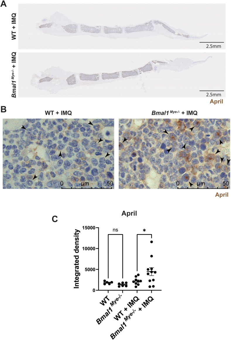
Figure 5.
Immunohistochemistry staining for the expression of April in mouse bone marrow. (A) Representative lower magnification (0.82×) images of IMQ-treated WT and Bmal1Mye−/− sternum bone marrow. April was stained with anti-mouse April antibody. (B) Higher-magnification (100×) images. April expressed in immature neutrophils (arrowhead) is increased in IMQ-treated Bmal1Mye−/− . (C) April levels in whole bone marrow were quantified with ImageJ and compared. WT; n=5, Bmal1Mye−/− ; n=5, WT + IMQ; n=9, Bmal1Mye−/− + IMQ; n=11. The statistical analysis was done using Mann–Whitney test. *p<0.05, ns, not significant.

