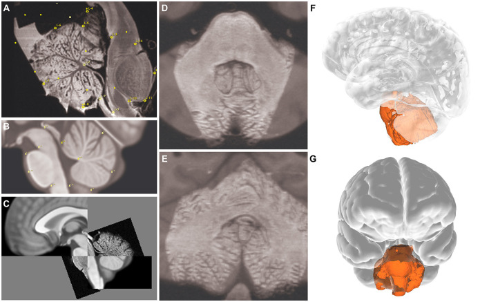
Figure 5.
Image planes showing landmarks (yellow marks) for the registration process. (A–C) Sagittal, (D) axial, and (E) coronal views. 3D reconstruction of the specimen overlaid on MNI space; (F) lateral and (G) anterior views. Used with permission from Barrow Neurological Institute, Phoenix, Arizona.

