
FIGURE 1.
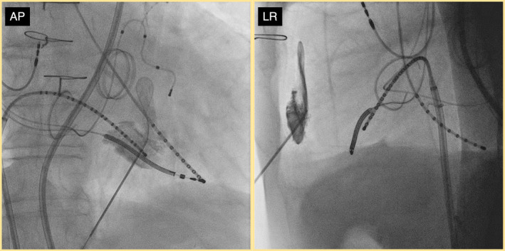
Fluoroscopic image showing the lack of contrast spread after attempted blunt dissection with the guide wire. This indicates significant pericardial adhesions preventing the guide wire from advancing. AP, Anterior–posterior view; LR, left–right view.