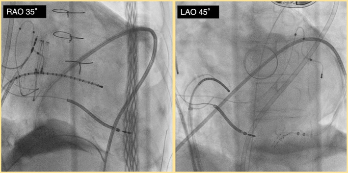
FIGURE 2.

Fluoroscopic image after successful dissection of the pericardial adhesions. After hydrodissection, dissection of the adhesions was performed using a catheter, and finally, the HD grid was inserted for mapping. RAO, right anterior oblique view; LAO, left anterior oblique view.
