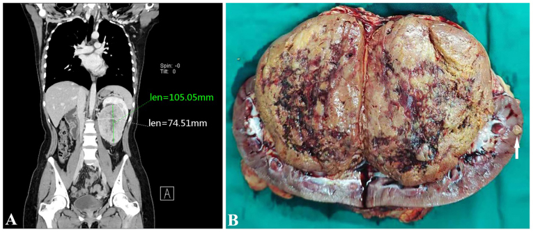
Figure 1.
CT examination and gross morphology of the present case. (A) Abdominal computerized tomography scan showed a 10.5×7.5 cm nodular mass with an uneven density in the left kidney. The boundary between the mass and the renal pelvis was not clear even after the left kidney was pushed upwards. (B) A brownish-yellow soft mass was present in the renal hilum and pelvis, with a clear boundary and shallow cut surface. Focal hemorrhage, cystic changes and small nodules (arrows) were seen in the renal parenchyma adjacent to the large mass. Len, length.

