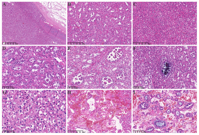
Figure 2.
Hematoxylin and eosin staining of the tumor tissue. (A) A pushing border surrounded the tumor (magnification, 4×). (B) The tumor was mainly composed of dense tubular and vesicular structures (magnification, 10×). (C) The tumor was arranged in solid vesicles (magnification, 10×). (D) Some tubules were dilated (magnification, 20×), with (E) tumor cells protruding into the lumen and forming characteristic annular tubular structures around eosinophilic hyaline bodies (magnification, 20×) or (F) calcifying in the lumen (magnification, 20×). (G) The morphology of the tumor cells was characterized by eosinophilic and flocculent cytoplasm (magnification, 40×). (H) The tumor stroma was rich in a thin-walled vascular network and some stroma showed signs of loose edema and bleeding (magnification, 10×). (I) Tumor cells in and around the stroma were diverse, including single cells rich in intracellular mucus, small to medium-sized round or dilated and twisted glandular ducts, and glandular lumen filled with gray-blue mucus magnification, (20×).

