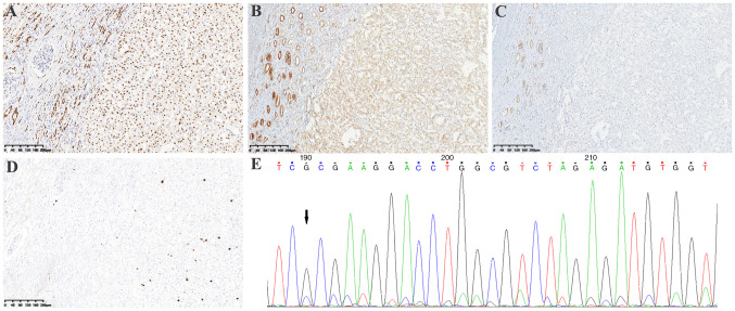
Figure 3.
Immunohistochemical staining of the tumor. Immunostaining showed tumor cells were (A) positive for PAX8 (magnification, 10×), (B) weakly positive for SDHA (the non-neoplastic renal tubules showed medium to strong positive staining; magnification, 10×), but (C) negative for SDHB (magnification, 10×). (D) Ki67 index was ~5% (magnification, 10×). (E) Sanger sequencing of SDHA: c.992_999dup (CCCCTGTC) (arrow). SDH, succinate dehydrogenase.

