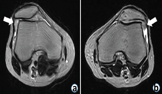
Figure 2.

Axial T2-weighted magnetic resonance images of the knee joints. (a) Synovial plica-like tissue was observed in the lateral patellofemoral joints of the right knee (white arrow). (b) Synovial plica-like tissue was observed in the lateral patellofemoral joints of the left knee (white arrow).
