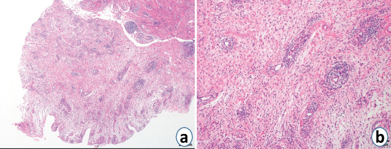
Figure 4.

Histopathological examination of tissue collected from the left lateral patellofemoral joint. (a) Granulation tissue with inflammatory cell infiltration and vascular hyperplasia were observed, but a tumor-like lesion with synovial proliferation was not detected (hematoxylin and eosin; ×40). (b) Inflammatory cell infiltration, vascular hyperplasia, and fibrous tissue were observed, consistent with findings for granulation tissue (hematoxylin and eosin; ×100).
