Figure 1.

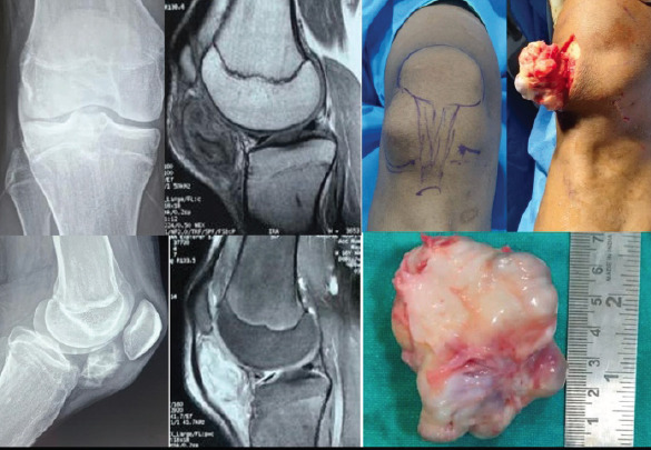
The radiological, clinical, and intraoperative images of the 16-year-old child with an infrapatellar fat pad calcified mass.

The radiological, clinical, and intraoperative images of the 16-year-old child with an infrapatellar fat pad calcified mass.