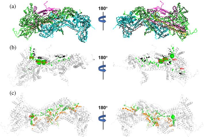
FIGURE 2.

Structural prediction of C2iso1,WT·WRC. (a) Superposition of C1iso1,WT·WRC X‐ray structure (black Cα‐trace) with our C2iso1,WT·WRC structural model. The five components of the complex (CYFIP2iso1, NCKAP1iso1, WAVE1, HSP300, and ABI2) are shown in green, cyan, magenta, yellow, and pink Ca‐traces, respectively. Cartoon representation of the (b) WAVE1/CYFIP2iso1 and (c) NCKAP1iso1/CYFIP2iso1 contact surfaces, colored in black/green and orange/green, respectively. The Cα atoms of the residues undergoing ASD‐associated mutations are labeled and shown as larger for adjacent residues (I664, E665 and D724, Q725) and smaller spheres for the others. The MR1 and MR2 loops of CYFIP2iso1 are also labeled. The C2iso2–3,WT·WRC complexes have similar configurations (Figures S3 and S4).
