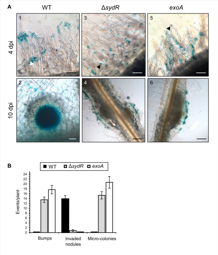
Fig 6.

ΔsydR mutant is defective in root infection. (A) Light microscopic images of M. truncatula roots inoculated with the WT (1, 2), ΔsydR (3, 4), and exoA (5, 6) strains expressing phemA:lacZ reporter fusion. β-Galactosidase activity (blue staining) was detected in (i) WT bacteria entrapped in root hairs (1), inside infection thread (white arrowhead) and nodule cells (2), and (ii) ΔsydR and exoA bacteria as micro-colonies mainly accumulated in RHCs (black arrowhead; 3, 5) and at the surface of bumps (4, 6). Scales: 50 µm (1, 2, 3, 5); 200 µm (4, 6). (B) Number of bumps, invaded nodules, and micro-colonies at 10 dpi. The values shown are the means ± SEM of three independent experiments. Significance compared to WT-inoculated roots was determined in Kruskal-Wallis and post hoc Conover-Iman tests with Benjamini-Hochberg correction (P < 0.05).
