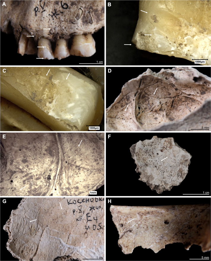
Fig 4. Kosenivka, selection of oral and pathological conditions.
A–E: Individual 5/6/+left maxilla. A: Teeth positions 23–26 (buccal view). Signs of periodontal inflammation (upper arrows) and examples of dental calculus accumulation (third arrow) and dental chipping (lower arrow) on the first premolar (tooth 24). B: First premolar (24, mesial view). Interproximal grooving with horizonal striations on the lingual surface of the root (upper arrow) and at the cemento–enamel junction (middle arrow). Larger chipping lesion (lower arrow). C: Canine (23, distal view). Interproximal grooving, same location as on the neighbouring premolar (see B), but less distinct. D, E: Signs of periosteal reaction on the left maxillary sinus (medio–superior view). Increased vessel impressions (D, upper arrow) and porosity, as well as uneven bone surface (D, lower arrow, E), indicating inflammatory processes. F: Individual 2, left temporal, fragment (endocranial view). Periosteal reaction indicated by porous new bone formation (arrow). G: Individual 5, frontal bone (endocranial view). Periosteal reaction indicated by tongue-like new bone formation and increased vessel impressions (arrows). H: Individual 5/6/+, frontal bone, right part, orbital roof (inferior view). Signs of cribra orbitalia (evidenced by porosity, see arrow). Illustration: K. Fuchs.

