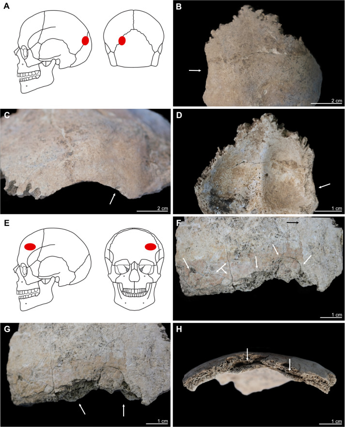
Fig 5. Kosenivka, selection of cases of perimortem cranial trauma, showing location and osteological details of the lesions.
A–D: Individual 1, occipital, left part. A: Location of the trauma, on the left posterior aspect of the cranium. B, C: Lamina externa (posterior and lateral views), showing an oval lesion (B, arrow) with a sharp rim and smaller punctual lesion (C, arrow). D: Lamina interna, typical uneven, terraced appearance and enlarged rim of the lesion (arrow). E–H: Individual 5, frontal bone, left part, anterior view. E: Location on the left anterior aspect of the cranium. F: Lamina externa (anterior view), showing multiple fracture lines and terrace-like lesion rims (arrows). G: Lamina externa (anterior–inferior view), showing the unevenly depressed rim of the lesion (arrows). H: View from the diploe, showing the deformation of the cranial bone, with splitting of the diploe (arrows). Illustration: K. Fuchs.

