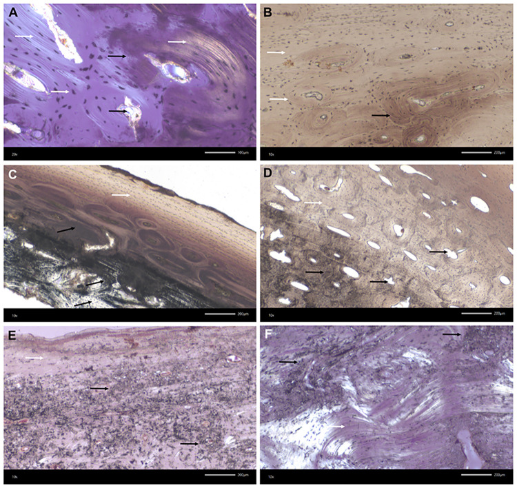
Fig 7. Kosenivka, transmitted light microscopy of bone thin-sections.
Microscopic features of preservation, fire impact and bioerosion in bone microstructure. A–D: Calcined bone. Black arrows indicate thermal impact, that is, calcined bone with discoloration by trapped carbon, burst Haversian canals, and reduced osteocyte distances due to shrinking. White arrows indicate well-preserved microstructures, such as osteons, osteocyte lacunae, circumferential lamellae, and collagen (A, yellow colour). A: Individual 1 (adult), right femur, proximal diaphysis, OHI of 5 (transversal section, polarised light). B: Individual 1, left parietal, cortical bone of lamina interna, OHI of 5 (vertical section, plain light). C: Individual 2 (younger child), left parietal, OHI of 5 (vertical section, plain light). D: Individual 3 (older child), left femur, diaphysis, OHI of 5 (transversal section, plain transmitted light). E, F: Unburnt bone with poorly preserved bone histomorphology. Strong impact of microbial attack visible by focal deconstruction (e.g., longitudinal tunnelling; black arrows; see Fig 17.5–17.7 in S1 Appendix) and a few well-preserved, original areas (white arrows). E, F: Individual 5/6/+ (adult), right humerus, distal epiphysis, OHI of 3 (transversal sections, polarised light). For more examples and age-related histomorphological features, see Fig 17 in S1 Appendix.

