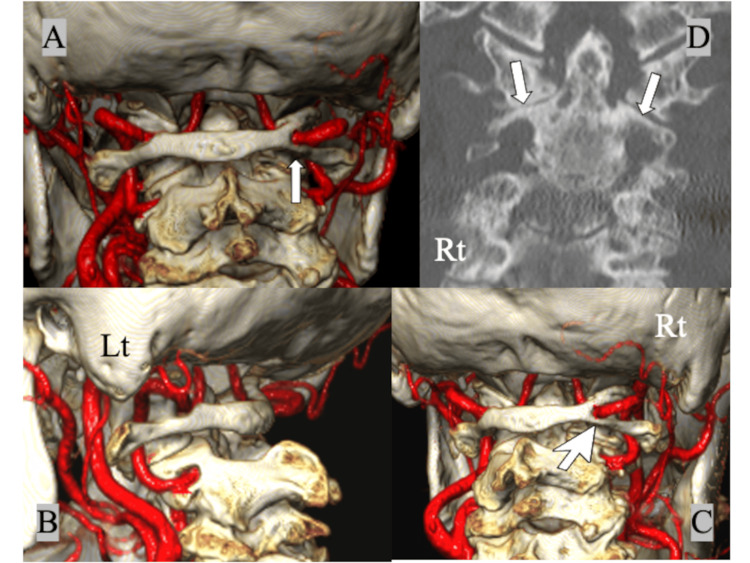
Figure 4. Preoperative 3D CTA showing (A) the bony bridge completely encircling the right VA on the right side (complete ponticulus posticus, white arrow), (B) a normal running course of the left VA on the VA groove, (C) a narrower posterior atlantal arch below the ponticulus posticus (white large-head arrow), and (D) bilateral high riding VAs with the thin C2 isthmus (white arrows).
3D: three dimensional; CTA: computed tomography angiography; VA: vertebral artery

