Figure 4.

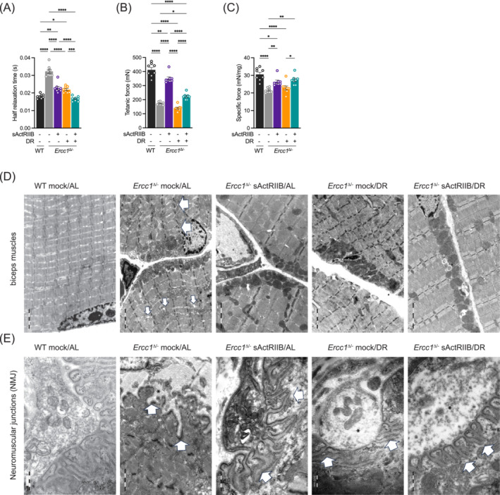
Impact of dual intervention on Ercc1 Δ/− muscle function and ultrastructure. Ex‐vivo assessment of (A) half relaxation time and (B) tetanic and (C) specific force. All muscles are from mice at 16 weeks of age. n = 8 WT Mock/AL, n = 7/8 Ercc1 Δ/− Mock/AL, n = 7/8, Ercc1 Δ/− sActRIIB/AL, n = 7/8, Ercc1 Δ/− Mock/DR, n = 7/8 Ercc1 Δ/− sActRIIB/DR. (D) sActRIIB but not dietary restriction prevents Ercc1 Δ/− muscle ultrastructural abnormalities. All transmission electron microscopy (TEM) images are from biceps muscle. In Ercc1 Δ/− mice, biceps muscles display disorganized, split sarcomeres of high variable width frequently containing a disrupted M‐Line. Moreover, the sarcoplasmic reticulum of these muscles was dilated. sActRIIB‐treatment alone as well as in combination with dietary restriction rescues this phenotype completely. In contrast, dietary restriction alone has no effect on any ultrastructural changes observed in Ercc1 Δ/− mice. Large arrows indicate disorganized sarcomeres and small arrows dilated sarcoplasmatic reticulum. (E) Neuromuscular junctions (NMJ) of biceps muscles in Ercc1 Δ/− mice display and obvious phenotype at the postsynaptic side. Namely in Ercc1 Δ/− mice the postsynaptic junctional folds of the basal lamina are almost completely absent or appear rudimentary. sActRIIB or dietary restriction could only partially rescue this phenotype, that is, the distance between the junctional foldings is still highly variable as well as their depth and width. Only double intervention by sActRIIB and dietary restriction fully restores the NMJ phenotype observed in Ercc1 Δ/− mice. *P < 0.05, **P < 0.01, ***P < 0.001, ****P < 0.0001.
