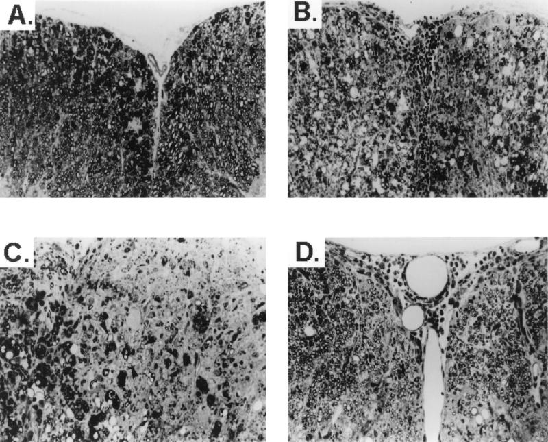
FIG. 2.
CTLA-4 Ig treatment leads to enhanced CNS myelin destruction and increased remyelination in TMEV-infected mice. (A) Section of spinal cord of hamster IgG-treated, TMEV-infected animal 60 days postinfection showing moderate myelin degeneration in both anterior columns. (B) Section from the spinal cord of a CTLA-4 Ig-treated mouse 63 days postinfection showing severe inflammation and demyelination in both anterior columns. (C) Section from a different area of the spinal cord of the CTLA-4 Ig-treated animal shown in panel B showing an area of active demyelination with numerous macrophages on the left and an area of chronic demyelination on the right. Arrowheads, axons surrounded by thin myelin, characteristic of remyelination. (D) Section from CTLA-4 Ig-treated animal 116 days postinfection shows mild residual inflammation, with both anterior columns showing extensive remyelination. Remyelinating cells are equally distributed between oligodendrocytes and Schwann cells. All sections are 1-μm-thick Epon-embedded sections stained with toluidine blue. Magnification, ×220.

