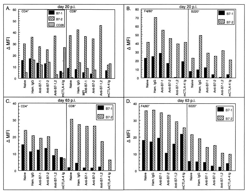
FIG. 7.
Phenotypic analysis of costimulatory molecule expression on splenic lymphocytes of TMEV-infected SJL mice treated with costimulatory molecule antagonists. Splenocytes from three representative animals per treatment group were harvested at 20 (A and B) and 63 days (C and D) postinfection and analyzed for cell surface expression of B7-1 and B7-2. For the analysis, cell gates were placed on CD4+, CD8+, F4/80+, or B220+ populations by histogram and these specific cell populations were analyzed for their levels of costimulatory molecule expression. Results are changes in mean fluorescent intensity (MFI) (MFI of cells stained with anti-B7 MAb − MFI of cells stained with isotype control antibody).

