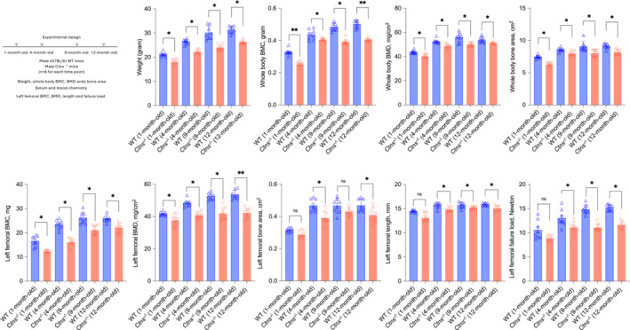
Figure 1.

Skeletal phenotype in Ctns −/− mice during the 12‐month study. All mice were fed ad libitum. Experimental design is listed. Mice were weighted to the nearest 0.1 g and then anaesthetized for in vivo scanning by dual‐energy X‐ray absorptiometry (DXA) to determine whole bone mineral content (BMC), bone mineral density (BMD) and bone area. The isolated left femora were assessed by DXA. Femoral BMC, BMD and bone area. Femoral length and load to failure were measured. Data are expressed as mean ± SEM. Results of Ctns −/− mice were compared with age‐matched WT mice. Ns, not significant, *P < 0.05, **P < 0.01.
