Fig. 2.
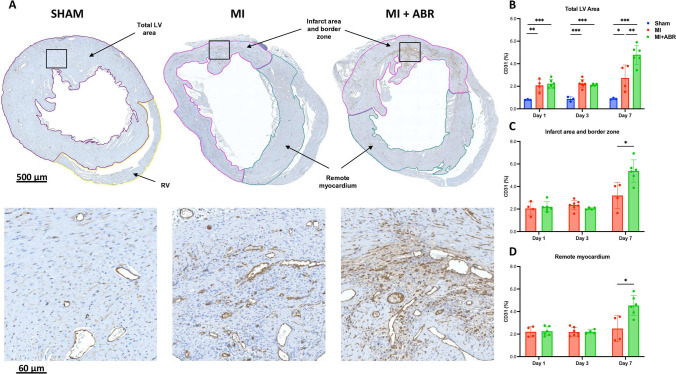
S100A8/A9 blockade induces enhanced neovascularization on day 7 post-MI. (A) CD31 staining in the three groups on day 7 post-MI. The images in the lower part of the figure are enlarged views of the myocardial areas marked by the black squares. The bar graphs show the CD31-positive area expressed as percent of the myocardial area at 1-day (Sham, n = 3; MI, n = 4; MI + ABR, n = 6; all females), 3-days (Sham, n = 3; MI, n = 7; MI + ABR, n = 4; all females) and 7-days post-MI (Sham, n = 3; MI, n = 4; MI + ABR, n = 6; all females) in the total LV area (B), infarct area and border zone (C) and remote myocardium (D). Data are presented as mean ± SD. *p < 0.05, **p < 0.01, ***p < 0.005. MI + ABR, mice with MI treated with ABR-238901 for up to 3 days post-MI

