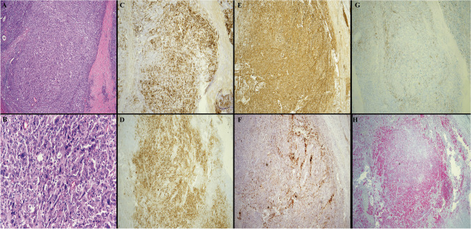
Fig. 1.
The composite picture represents parotid gland involvement by follicular dendritic cell sarcoma. A H&E stain, low-power magnification (4 ×) of the sarcoma cells with rare unremarkable glands noted. B H&E stain, high-power magnification (40 ×) showing the large sarcoma cells, mostly with one centrally placed nucleoli and open chromatin. Scattered large highly atypical malignant cells are noted. Few interspersed small lymphocytes are also seen. C CD21 expression by the sarcoma cells (4 ×). D CD23 expression (4 ×). E Clusterin expression (4 ×). F CXCL13 expression (4 ×). G CD35 partial expression (4 ×). H PRAME expression (4 ×)

