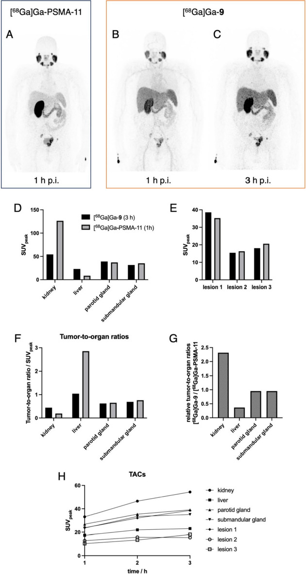
Fig. 3.
PET imaging of a 60-year old male prostate carcinoma patient with multiple lymph node metastases. A Coronal MIP PET image at 1 h p.i. with 113 MBq [68Ga]Ga-PSMA-11. B + C Coronal MIP PET images at 1 and 3 h using 107 MBq [68Ga]Ga-9, respectively. Comparison of SUVpeak of [68Ga]Ga-PSMA-11 at 1 h p.i. vs. [68Ga]Ga-9 at 3 h p.i. D in healthy organs and E in the three target lesions (1 × prostate, 2 × lymph node metastases). F Comparison of corresponding tumor-to-organ ratios. G Relative ratio of tumor-to-organ ratios between [68Ga]Ga-PSMA-11 and [68Ga]Ga-9. H Time activity curves (TACs) for [68Ga]Ga-9 at 1, 2, and 3 h p.i

