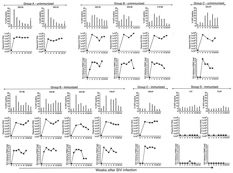
FIG. 1.
Longitudinal analysis of lymphocyte proliferation, plasma SIV RNA, and infectious SIV load in PBMC in individual rhesus macaques inoculated with pathogenic SIV. Data for seven animals immunized with an HSV recombinant expressing SIV proteins prior to SIV inoculation are shown in the bottom half of the figure. Data for six animals that were SIV naive prior to SIV inoculation (includes one animal, 285.95, that received a control HSV recombinant) are shown in the top panel. The animals were grouped on the basis of time of appearance of peak increase in Ki-67 expression in total lymphocytes in peripheral blood following SIV inoculation. This occurred at 1 week in group A, at 2 weeks in group B, and at 4 weeks after SIV inoculation in group C animals. In group D animals, there was no change in Ki-67 expression following SIV inoculation. Infectious SIV loads in PBMC were not available for the two group A animals.

