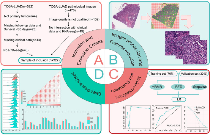
Figure 1.
Data analysis workflow. (A) Inclusion criteria for the study. (B) Processing of the pathological images. (C) The process of establishing and evaluating the pathomics model. (D) The relevant mechanisms explored. TGCA, The Cancer Genome Atlas; LUAD, lung adenocarcinoma; mRMR, max-relevance and min-redundancy; RFE, recursive feature elimination; LR, logistic regression; ROC, receiver operating characteristic; AUC, area under the curve; DCA, decision curve analysis; PS, pathomics score.

