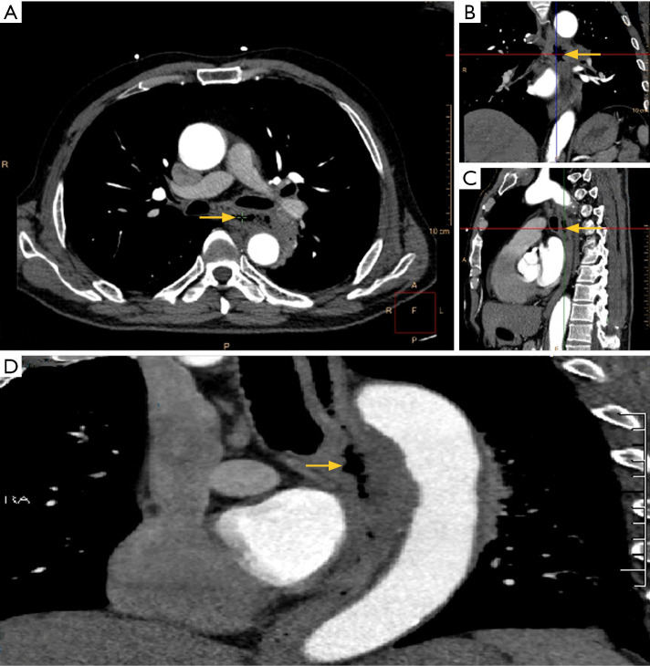
Figure 3.
Computed tomography observation of the perforation from the horizontal plane (A), coronal plane (B), and sagittal plane (C). Curved planar reconstruction (D) clearly demonstrated the discontinuity of the esophageal wall, vividly depicting the ruptured area. Yellow arrows in all plans indicate the discontinuity of the esophageal wall.

