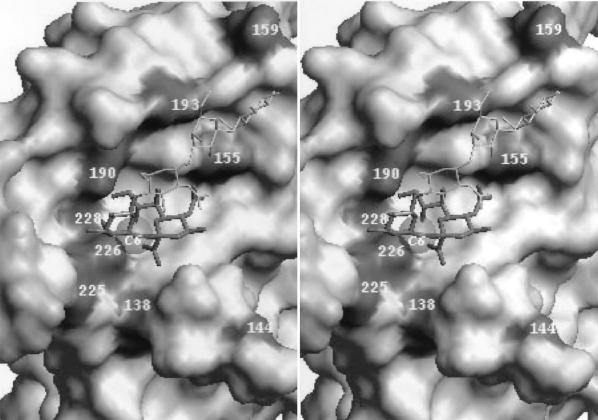
FIG. 3.
Positions of amino acids in the HA receptor-binding site that differ between early epidemic human and swine viruses and their closely related avian counterparts (stereo view). The figure is based on the crystallographic model of the X31 HA complex with the Neu5Acα2-6Gal-containing receptor analog LSTc (Neu5Acα2-6Galβ1-4GlcNAcβ1-3Galβ1-4Glc) (10). The solvent-accessible molecular surface of the protein is shown. The sialic acid residue and penultimate galactose ring of LSTc are displayed as thick stick bonds; the rest of the molecule is shown as a thin white line. The gray transparent sphere (C6) in close proximity to amino acid 226 represents the van der Waals surface of the C6′-carbon atom of Gal. The figure was generated with WebLab ViewerPro 3.10 (Molecular Simulations, Inc., San Diego, Calif.).

