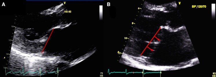
Figure 1.
The mitral valve with and without mitral valve prolapse. Transthoracic echocardiographic long-axis view in end-systole zoomed in on the mitral valve with (A) the location of both mitral leaflets within <2 mm under the plane of the mitral annulus (red line) refutes the diagnosis of mitral valve prolapse. (B) A displacement of both leaflets >2 mm (red arrow) above the plane of the annulus (red line) defines mitral valve prolapse. Note the absence of detachment of the posterior leaflet from the left ventricular myocardium (no mitral annular disjunction).

