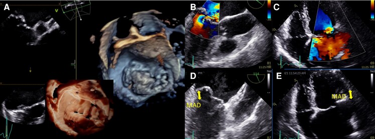
Figure 3.
Mitral valve prolapse and mitral annular disjunction (MAD) on transthoracic echocardiography (TTE) and transoesophageal echocardiography (TEE). (A) TEE 3D view, displaying a large prolapse of P2 precisely assessed by ‘photo-realistic’ tools now available. Severe mitral regurgitation is associated as shown on 120° TEE (B) and TTE apical four-chamber views (C), with severe left atrial dilatation. A 10 mm MAD is diagnosed both on these TEE (D) and TTE (E) views.

