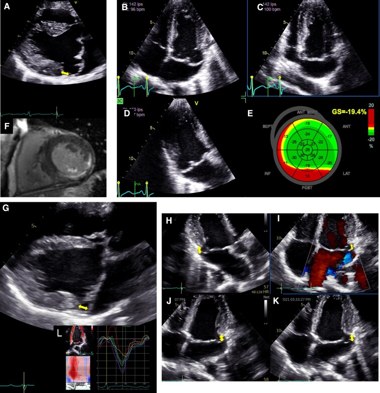
Figure 7.
Left ventricular mechanical dispersion associated with mitral annular disjunction (MAD). First panel: Transthoracic echocardiographic long-axis view in end-systole (A) displaying mitral valve prolapse with MAD (yellow arrow). Transthoracic echocardiographic apical four-chamber (B) three-chamber (C) and two-chamber (D) views for the measurement of global longitudinal strain (E). Note the disjunction inducing mechanical dispersion and post-systolic shortening of the posterior-basal segment of the left ventricle (in red). The patient has been seen for ventricular premature complexes. (F) The disjunction is associated with fibrosis in the basal inferolateral segments of the left ventricle and mild mitral regurgitation. Second panel: Transthoracic echocardiographic long-axis view in end-systole (G) displaying mitral valve prolapse with MAD (yellow arrow). Note here the disjunction involving all the insertion of the posterior leaflet on the annulus on transthoracic echocardiographic apical three-chamber (H) and four-chamber views (I, J, K). The depth of the disjunction is only mild yet the circumferential extension is extensive. (L) It leads to mechanical dispersion, post-systolic shortening of the posterior-basal segment of the left ventricle. The patient has been seen for palpitations and > 10% ventricular premature complexes.

