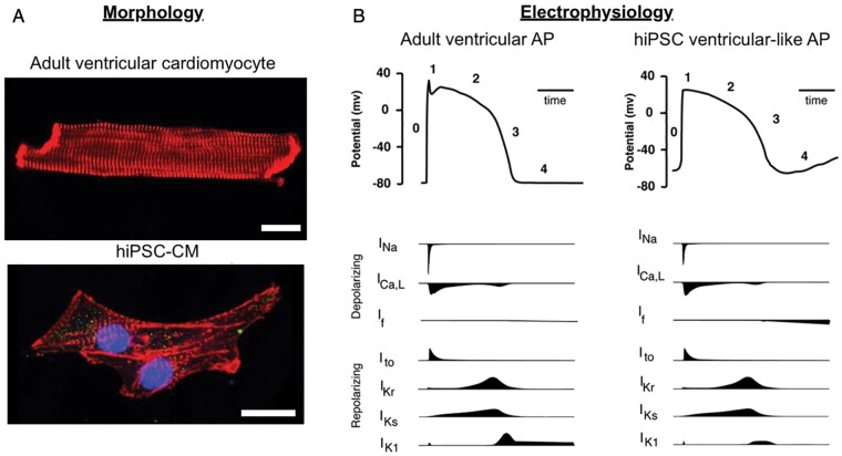
Figure 3.
Comparison of morphological and electrophysiological features of adult ventricular cardiomyocytes and human-induced pluripotent stem cell (hiPSC)-derived cardiomyocytes. (A) Microscopic image of an adult cardiomyocyte from mouse stained with anti-alpha-actinin (red) and a hiPSC-derived cardiomyocyte stained with anti-alpha-actinin (red), DAPI (blue), and Nav1.5 (green). Scale bars are 20 µm. (B) Schematic of ventricular action potentials (APs) during rapid upstroke (Phase 0), early repolarization (1), plateau (2), late repolarization (3), and diastole (4). The ionic currents involved are shown below the AP traces.

