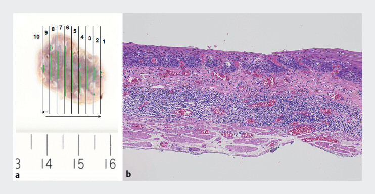
Fig. 3.
Macroscopic and histopathological images of the resected specimen. a Macroscopic image of the specimen. b Histopathological image of the specimen. The pathological diagnosis was esophageal squamous cell carcinoma in the lamina propria mucosae with no lymphovascular invasion and negative margins.

