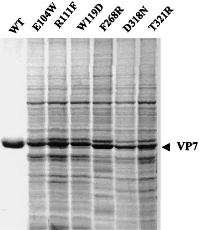
FIG. 3.
Expression of recombinant VP7 mutants. Sf cells were infected with each recombinant virus, and the infected-cell lysates were analyzed by SDS–10% PAGE and stained with Coomassie brilliant blue. Each mutant protein is indicated. The sizes of the expressed proteins are compared with the purified wild-type VP7 protein as indicated (WT). The position of VP7 is shown by an arrow.

