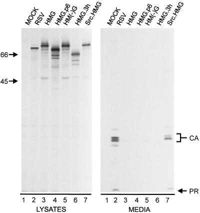
FIG. 3.
Release properties of modified forms of HMG. The indicated constructs were transfected into duplicate plates of COS-1 cells. The right panel (media) was from one set of plates that were treated as described in the legend to Fig. 2. Gag proteins in the left panel (lysates) were collected and visualized as for Fig. 2, but were from the second set of plates that had been labeled for only 5 min.

