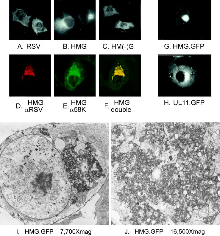
FIG. 4.
Microscopic analyses of HMG-expressing cells. COS-1 cells were transfected with the indicated constructs. Cells in panels A to F were analyzed by immunofluorescence. In panels A to C, antibodies specific for RSV Gag were used. Cells in panels D to F were double labeled with a mixture of rabbit antibodies against Gag and mouse antibodies against the Golgi 58K protein, which were detected by using a mixture of goat anti-rabbit antibodies conjugated to TRITC and goat anti-mouse antibodies conjugated to FITC. Panels D and E are the same field viewed by confocal microscopy with the appropriate wavelength to excite TRITC (D) or FITC (E) and were digitally combined to provide the image in panel F. Cells transfected with GFP constructs in panels G and H were viewed by light microscopy without fixing or staining. Cells in panels I and J were analyzed by standard electron microscopy.

