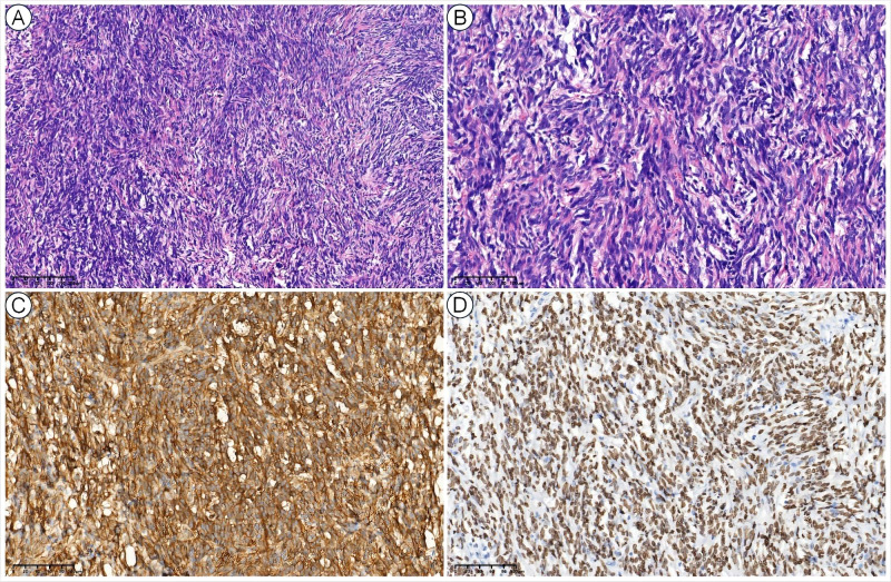
Figure 2.
Histopathological views of SFTP. The low-power (10×) hematoxylin and eosin (H&E) view showed a patternless pattern (A), and the high-power (40×) H&E view showed enlarged spindle cells with a variable proportion of collagenous stroma (B). The tumor cells were also positive for CD34 (C) and STAT6 (D).

