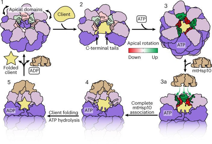
Fig. 6. Model of conformational changes in the client-engaged mtHsp60 reaction cycle.

State 1, ADs (pink) of mtHsp60apo heptamers are flexible and exhibit modest rotation about the apical–intermediate hinge, denoted by coloration of helices H and I. State 2, Client binding to mtHsp60apo preserves AD asymmetry, and client can localize to multiple depths of the heptamer, facilitated by mtHsp60 ADs and the flexible C-terminal tails. State 3, ATP binding induces the dimerization of heptamers through the equatorial domains and a more-pronounced AD asymmetry in an alternating up/down arrangement. ADs in ‘down’ protomers (red) contact client, whereas those in ‘up’ protomers (green) are competent to bind mtHsp10. State 3a, mtHsp10 initially binds the mtHsp60 heptamer using the three upward-facing ADs; all ADs then transition to the conformation observed in the mtHsp10-bound complex (state 4). After ATP hydrolysis and client folding (state 5), client, mtHsp10 and ADP are released, and the double-ring complex disassociates into heptamers.
