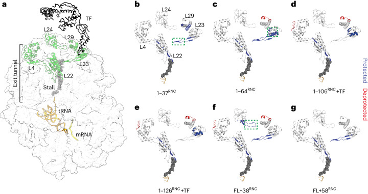
Fig. 6. Effect of nascent DHFR on ribosomal proteins.
a, Side view of the 70S ribosome, highlighting ribosomal proteins that line the exit tunnel and vestibule (PDB 3JBU). TF is placed onto the structure based on a previous publication56. The structure contains the 17-aa wild-type SecM stall-inducing sequence, although to match the length of the sequence used in this study only eight amino acids are shown. b–g, HDX analysis of ribosomal proteins in RNCs. Peptides that are protected from HDX in each RNC relative to empty ribosomes, at any deuteration time point, are colored blue. Deprotected peptides are colored red. The tunnel-facing loop of L23 is indicated with a green dashed rectangle in b, the TF docking site is indicated with a green dashed rectangle in c and the exposed loop of L24 is indicated with a green dashed rectangle in f. See also Supplementary Data 1.

