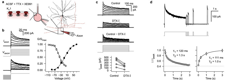
Fig. 2. Axons of human layer 2 & 3 pyramidal neurons contain Kv1 potassium channels that show slow inactivation in the subthreshold voltage range.
a Reconstruction of biocytin labeled pyramidal neuron (dendrite black, axon red) and schematic of ‘whole-bleb’ recording configuration (see Methods: Axonal voltage-clamp recordings). b Left, K+-currents in response to activation and steady-state inactivation voltage-clamp protocols (see Methods). Right, normalized K+-conductance as a function of test voltage (black data points, activation protocol, n = 9 axonal recordings) or conditioning voltage (white data points, inactivation protocol, n = 5 axonal recordings). Data is displayed as mean ± s.e.m. c K+-currents in response to voltage steps during control period, after local puff-application of dendrotoxin-I (DTX-I), as well as subtraction of the two conditions. Bottom, summary plot showing peak currents in response to +70 mV test pulses before and after DTX-I application (n = 5 axonal recordings). d K+-currents in response to a protocol to determine inactivation and recovery from inactivation kinetics (see Methods). Bottom, summary plots displaying mean ± s.e.m of normalized current amplitudes (n = 5 axonal recordings). Lines correspond to double exponential fits (time constants are shown as insets). ACSF, artificial cerebrospinal fluid; TTX, tetrodotoxin. Source data are provided as a Source Data file.

