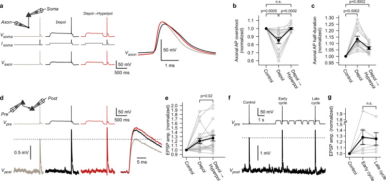
Fig. 4. Sequences of presynaptic de- and hyperpolarizations recover axonal action potential amplitude and further increase synaptic transmission.
a Exemplary somato-axonal recording. Somatic current injections (Isoma) were used to induce ‘Depol’ and ‘Depol→Hyperpol’ conditions. Right, magnified axonal action potentials (AP). Note the decrease and rescue of axonal AP overshoot in the ‘Depol’ and ‘Depol→Hyperpol’ conditions, respectively. b, c Summary graphs showing relative changes of the axonal AP overshoot and half-duration (n = 13 somato-axonal recordings; two-sided Wilcoxon signed-rank tests; error bars show mean ± s.e.m). d Exemplary paired recording of synaptically connected pyramidal neurons. Excitatory postsynaptic potentials (EPSP) were averaged over multiple trials and are shown on a finer timescale on the right. e Summary graph showing relative changes of EPSP amplitudes (n = 21 paired recordings; two-sided Wilcoxon signed-rank test; error bars show mean ± s.e.m). f Exemplary paired recording. ‘Control’ condition was compared to early and late cycles of multiple de- and hyperpolarizations. g Summary graph showing relative changes of the average EPSP amplitudes (n = 7 paired recordings; two-sided Wilcoxon signed-rank test; error bars show mean ± s.e.m). Source data are provided as a Source Data file.

