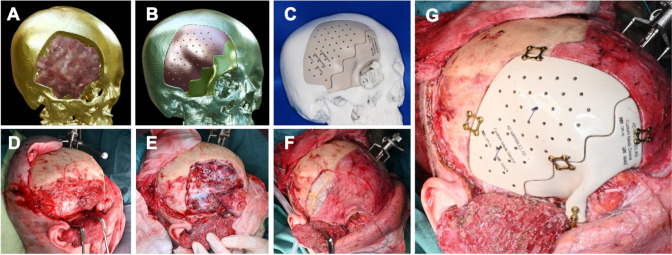
Fig. 2.
The workflow from virtual planning to implant insertion is shown in Fig. 2. 3-dimensional virtual planning shows tumor extention (red mass) and preplanned bony resection margins (A), the individually designed implants are previewed (B) and both, a template of the skull after craniectomy and the implants are 3D printed and used for preoperative and intraoperative planning (C). Intraoperative images show the marks for craniotomy on the skull (D), the skull after craniectomy with a thoroughly decompressed orbit (E) and dural closure with pericranium and Tutopatch (Biomedica, Milan, Italy) dural replacement (F). The final result shows optimal positioning of the two implants and fixation with titanium micro plates (G) (Medartis, Basel, Switzerland)

