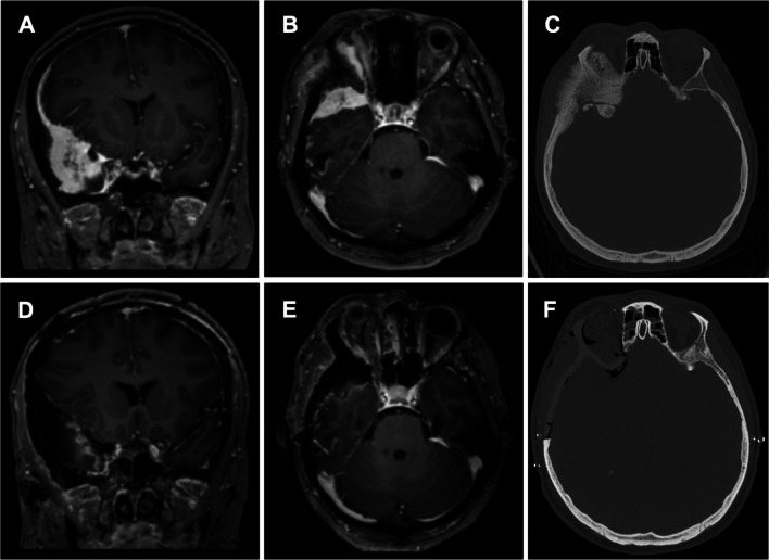
Fig. 4.
(patient #1) MRI of the head T1 sequences with contrast medium and thin-sliced CT scan of the head. A contrast medium enhancing lesion is seen at the right temporal pole (A) with infiltration of the orbital contents (B). In the bone window CT scan invasive growth into the frontal bone, the sphenoid wing, the zygomatic process and the lateral orbit is shown (C). Postoperative MRI scan with contrast medium reveals good tumor removal in the coronar (D) as well as in the axial view (E). CT scan of the head on the following day showed an optimal positioning of the PEEK implant with a good symmetrical result (F)

